-
![]()
![]()
Combining grain boundary diffusion and segmentation: A novel production route for resource-efficient Nd–Fe–B magnets
2026/05/28
New publication
-
![]()
![]()
New ACS Omega publication on anomalous Nernst effect in Fe4–xGexN thin films
2026/02/11
-
![]()
![]()
FM group again part of the Erfinderlabor
2026/02/10
40th Erfinderlabor – High Performance Materials and Recycling – A Contradiction?
-
![(a) Hysteresis and initial magnetization curves for the low and high Hc samples. (b) 3D atom probe tomography (APT) data comparing low Hc and high Hc samples. Top: 3D APT reconstructions of low Hc and high Hc samples, respectively, with Co (dark blue), Ce (red), Cu(light blue); Middle: 2D Cu concentration maps from 10 nm thick slices; Bottom: 1D concentration profiles extracted from cylinders (5 nm diameter, 100 nm length). (c) Magnetic imaging of Ce(Co0.8Cu0.2)5.4 high Hc sample: Fresnel Lorentz Transmition Electron Microscopy image, showing black and white contrast at the positions of zigzag domain walls (DW), nucleated under applied field B = 1T. The image is recorded in magnetic-field-free conditions at a defocus of 1 mm. (d) Magnetic induction map of a DW region indicated by a yellow dotted square in (c). Arrows and colours indicate projected in-plane magnetic field directions. (e) Schematic representations of the coercivity mechanism in Ce(Co0.8Cu0.2)5.4 obtained by micromagnetic simulation: High Hc sample shows pronounced chemical contrast and preferred orientation of Cu-poor cylindrical cells leads to strong pinning at Cu-rich cell boundaries. Low Hc sample demonstrates weaker compositional contrast and random cell orientation reduce pinning effectiveness. The DW (dashed line) propagates more easily through the less segregated structure.]()
![(a) Hysteresis and initial magnetization curves for the low and high Hc samples. (b) 3D atom probe tomography (APT) data comparing low Hc and high Hc samples. Top: 3D APT reconstructions of low Hc and high Hc samples, respectively, with Co (dark blue), Ce (red), Cu(light blue); Middle: 2D Cu concentration maps from 10 nm thick slices; Bottom: 1D concentration profiles extracted from cylinders (5 nm diameter, 100 nm length). (c) Magnetic imaging of Ce(Co0.8Cu0.2)5.4 high Hc sample: Fresnel Lorentz Transmition Electron Microscopy image, showing black and white contrast at the positions of zigzag domain walls (DW), nucleated under applied field B = 1T. The image is recorded in magnetic-field-free conditions at a defocus of 1 mm. (d) Magnetic induction map of a DW region indicated by a yellow dotted square in (c). Arrows and colours indicate projected in-plane magnetic field directions. (e) Schematic representations of the coercivity mechanism in Ce(Co0.8Cu0.2)5.4 obtained by micromagnetic simulation: High Hc sample shows pronounced chemical contrast and preferred orientation of Cu-poor cylindrical cells leads to strong pinning at Cu-rich cell boundaries. Low Hc sample demonstrates weaker compositional contrast and random cell orientation reduce pinning effectiveness. The DW (dashed line) propagates more easily through the less segregated structure.]()
New Acta Materialia publication
2026/02/02
Direct observation of nanoscale pinning centers in Ce(Co0.8Cu0.2)5.4 permanent magnets
-
![a) Microstructure of the magnet obtained by electron microscopy. (b) Optical Kerr microscopy image, the black areas correspond to the demagnetized part of the magnet. (c,d,e) High-resolution transmission electron microscopy images showing nanostructure of the magnet. (f) Electron holography, different colours correspond to different orientation of magnetization.]()
![a) Microstructure of the magnet obtained by electron microscopy. (b) Optical Kerr microscopy image, the black areas correspond to the demagnetized part of the magnet. (c,d,e) High-resolution transmission electron microscopy images showing nanostructure of the magnet. (f) Electron holography, different colours correspond to different orientation of magnetization.]()
How defects make permanent magnets even more efficient
2026/01/28
TU researchers contribute to a publication in Nature Communications
-
![]()
![]()
New Advanced Materials Publication with National Institute for Material Science (NIMS – Japan)
2026/01/22
-
![]()
![]()
PhD Defense Konrad Opelt
2025/12/10
-
![]()
![]()
Dean’s Prize for Excellence in Research
2025/12/05
-
![]()
![]()
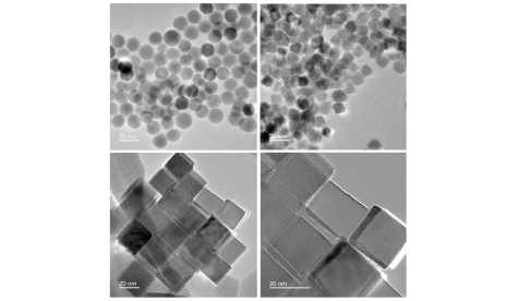
The influence of the shape of magnetic nanoparticles on magnetic hyperthermia and cellular internalisation
2025/11/14
Published in Journal of Alloys and Compounds
-
![]()
![]()
Media review: TU Professor Gutfleisch on Rare Earth Elements
2025/11/06
Why Rare Earth Magnets Are So Hard to Replace
Archive