

Materials in Radiation Fields
In a number of applications, such as in nuclear facilities, particle accelerators and in space, materials are exposed to energetic ionizing radiation. The irradiation may lead to a degradation of the material properties.
Polymers are particularly sensitive in this regard. In a collaboration with GSI , polyimide and polyepoxide, which are components of superconducting beam guiding magnets, are irradiated with relativistic heavy ions and characterized for their properties, such as network degradation and electrical conductivity.
Another material is polycrystalline graphite, which is being used as the target wheel and as the beam dump.
Studies on inorganic scintillators for high-current diagnosis of heavy ion irradiation
Scintillation screens are used at particle accelerator facilities for beam diagnostics in order to obtain a two-dimensional beam profile and thus to determine the position, shape and intensity distribution of the ion beam. For this purpose, the phosphor screen is inserted into the ion beam, excited by it to scintillation, and the emitted light is registered by a suitable detector (e.g. a CCD camera). In addition to the direct imaging of the beam, scintillation screens are often used in other diagnostic tools, such as in the so-called pepper-pot device for determining the brightness of the beam or as an imaging element behind multi-channel plates (MCP).
In general, the scintillation screen will be exposed directly to the primary ion beam, i.e. the used materials have to be applicable for extreme conditions such as high beam intensities and energies. The resulting radiation damage leads to the rapid degradation of the material and can distort the imaging properties of the scintillation screens.
Thus, the scintillation properties of ceramic screens used for high-current ion beam imaging are studied. The screens are tested under heavy-ion irradiation with varying beam parameters (projectile ion, energy, energy loss dE/dx, particle fluence, target temperature). Various inorganic materials were investigated in two sub-projects:
Low-energy: The experiments were performed at two accelerator facilities, the UNILAC at GSI Helmholtz Centre for Heavy Ion Research GmbH in Darmstadt and at 6 MV Tandetron accelerator of Helmholtz Centre Dresden-Rossendorf (HZDR) in Dresden with particle energies ranging from 0.5 to 11.4 MeV/u.
High-energy: The irradiation experiments were done at the heavy ion synchrotron SIS18 at GSI Helmholtz Centre for Heavy Ion Research GmbH with energies of several hundred MeV/u.
The experiments focus not only on conventionally used scintillators (for example, P43, P46, Al2O3:Cr), but also on radiation resistant ceramics (for example, Al2O3, ZrO2). The imaging properties of the materials (light yield, beam width and higher statistical moments) were compared. The image of different ion beam pulses was taken with a digital CCD camera and evaluated individually. In the low-energy range, in general a material degradation can be observed resulting in a decreasing light yield as a function of the irradiation time. This is attributed to the massive generation of radiation damage (vacancies, interstitials etc.). Moreover, the received beam width depends on the scintillation material.
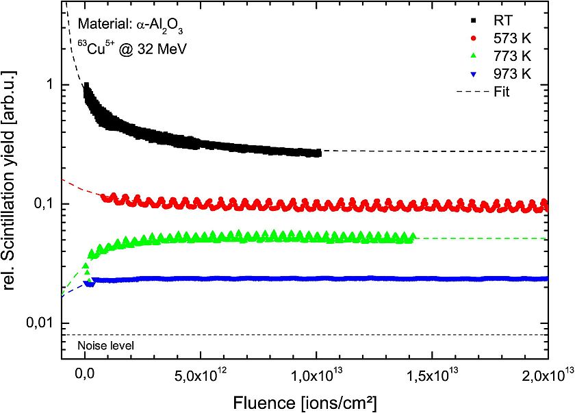
In addition, both, light yield and beam width depend on the screen temperature, which increases by the ion irradiation. With increasing temperature, a decrease in light yield is recorded caused by thermal quenching. However, it could be shown that by a specific thermal treatment of the materials, the further degradation of the light yield can be decelerated or even stopped (Figure 1). This is attributed to diffusion driven annealing processes of the generated radiation defects. Thus, the annealing of the materials is an effective tool to extend the lifetime of the screens.
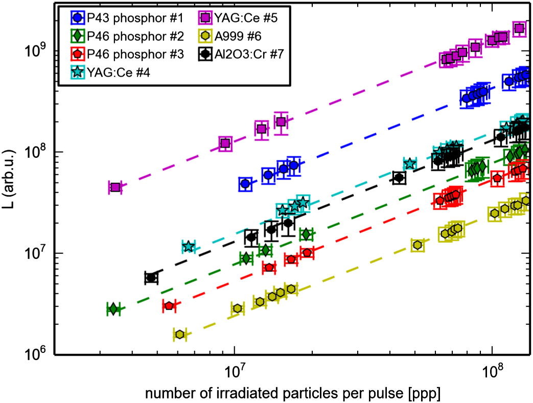
For the irradiation experiments performed in the high-energy range the degradation of the screens outlined above does not occur. Thus beam diagnostics can resort to conventionally used scintillators. The most efficient conversion of energy into light was measured with a phosphor screen P43. The highest light output in total was measured by a cerium-doped YAG crystal. However, this crystal showed the greatest deviations in the imaging of the beam profile (Figure 2). Both effects are due to the screen thickness of about 1 mm. A decrease in light output as a function of irradiation time was barely recordable if at all. The reason for this behavior is the less intensive production of radiation damage, since only a fraction of the projectile energy is deposited directly in the material (Bragg peak). However, a drop in light output can be detected with increasing atomic number of the projectile. This may possibly be explained by the non-proportionality of scintillators.