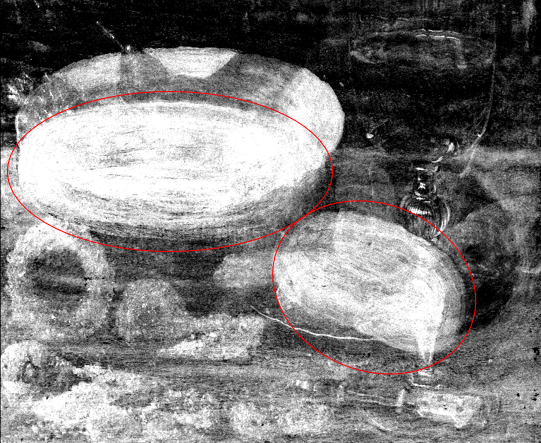
During a MA-XRF experiment, the painting is excited by an X-ray milli-beam to emit X-ray fluorescence radiation. Via the energy of the recorded fluorescence radiation the elements present in the analyzed spot can be identified. By moving the beam relative to the painting, the surface is scanned pixel by pixel. In a next step, the resulting digital data is converted into one or more elemental distribution images by means of software, allowing to study the distribution of various pigments on or beneath the entire paint surface in a visual manner, and this in a non-contact/non-destructive way.
MA-XRF analysis on paintings are usually carried out to support an ongoing conservation treatment with insights into the artist's materials, techniques and degradation mechanisms. Nevertheless, for several master’s such as Van Eyck, Rembrandt or Vermeer the aim is to obtain a significant corpus of data that allows to characterize the studio practice of these artists. The elemental distribution images are also able to reveal and assess the extent of hidden paintings.
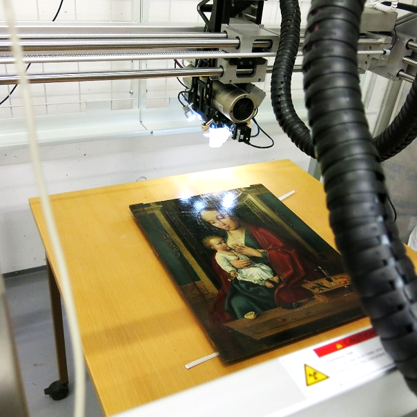
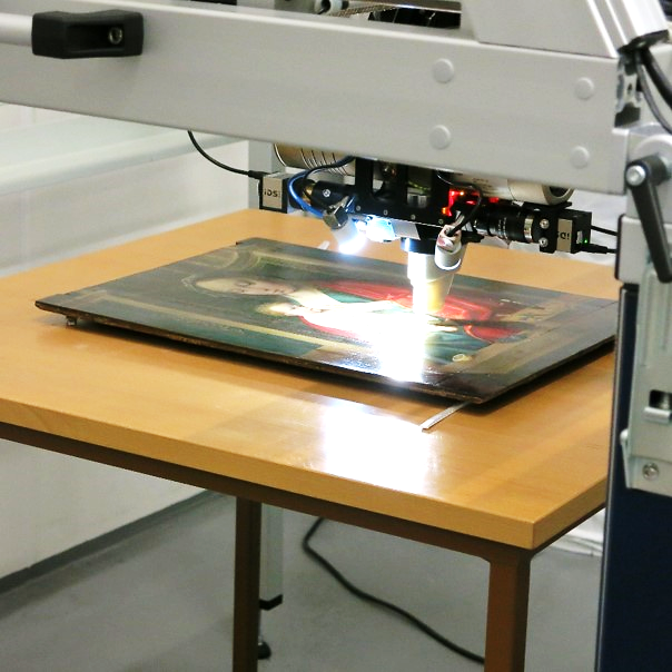
MA-XRF
Since its development in 2008, macroscopic X-ray fluorescence scanning (MA-XRF) has allowed to gain insight about the composition and build-up of polychrome works of art by means of elemental distribution maps, obtained in situ through non-destructive analysis. Such scanning instrument provides chemical information from the entire object (both on and below the surface) while the ensuing analytical results are presented in the form of an image
During a MA-XRF experiment, the painting is excited by an X-ray milli-beam to emit X-ray fluorescence radiation. Via the energy of the recorded fluorescence radiation the elements present in the analyzed spot can be identified. By moving the beam relative to the painting, the surface is scanned pixel by pixel. In a next step, the resulting digital data is converted into one or more elemental distribution images by means of software, allowing to study the distribution of various pigments on or beneath the entire paint surface in a visual manner, and this in a non-contact/non-destructive way.
MA-XRF analysis on paintings are usually carried out to support an ongoing conservation treatment with insights into the artist's materials, techniques and degradation mechanisms. Nevertheless, for several master’s such as Van Eyck, Rembrandt or Vermeer the aim is to obtain a significant corpus of data that allows to characterize the studio practice of these artists. The elemental distribution images are also able to reveal and assess the extent of hidden paintings.
The MA-XRF portable equipment employed facilitates collaboration with curators, scholars, and private collectors, to assist with questions of process and authenticity, using scientific evidence considered together with connoisseurship, provenance, and art historical research.
The MA-XRF portable equipment employed facilitates collaboration with curators, scholars, and private collectors, to assist with questions of process and authenticity, using scientific evidence considered together with connoisseurship, provenance, and art historical research.