Functional Materials

Science Gallery
Welcome to our Functional Materials “Magnetism as Art Gallery”
Besides their fascinating functionalities, we also want to showcase the beauty and complexity of magnetism and magnetic materials. When documenting our research, we sometimes discover hidden and surprising aesthetic structures, textures and shapes. You are welcome to use these images and videos in your works and presentations, and acknowledge our ©Functional Materials Group at TU Darmstadt, thanks and enjoy.
Images
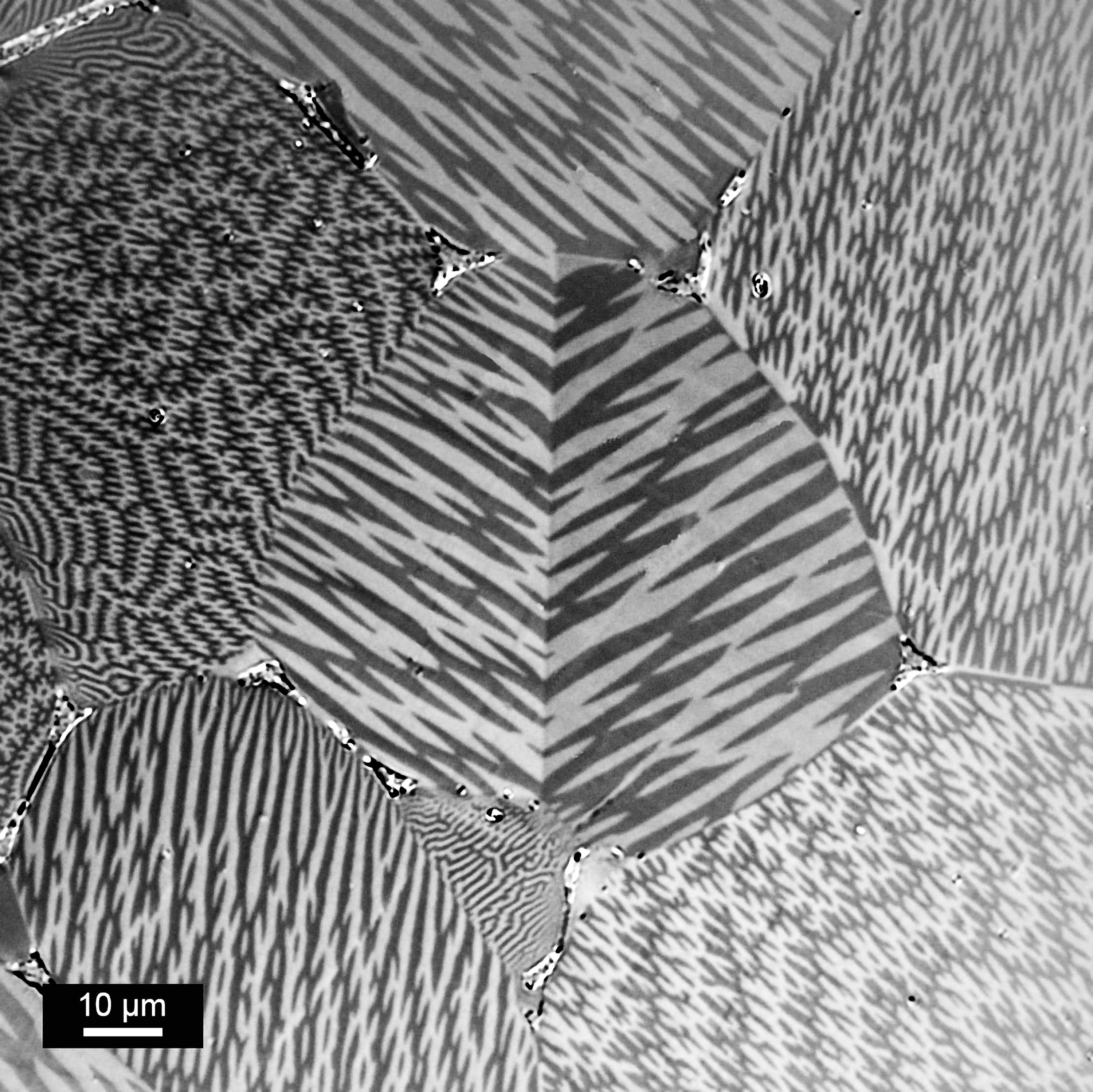
Kerr microscopy from a commercial SmZrCoFeCu alloy after induction melting.
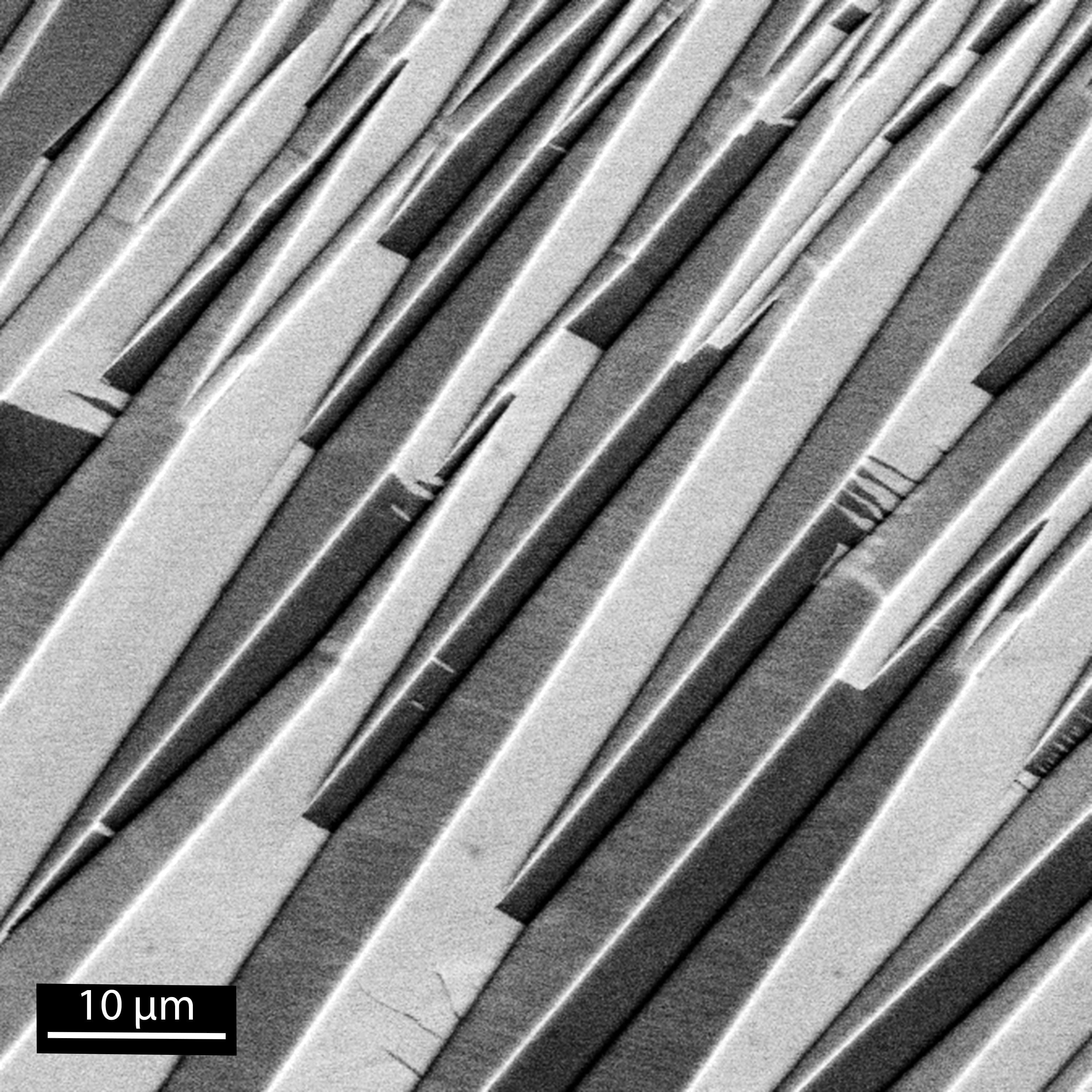
Fe3Sn single crystal (hexagonal structure) grown using reactive flux technique.
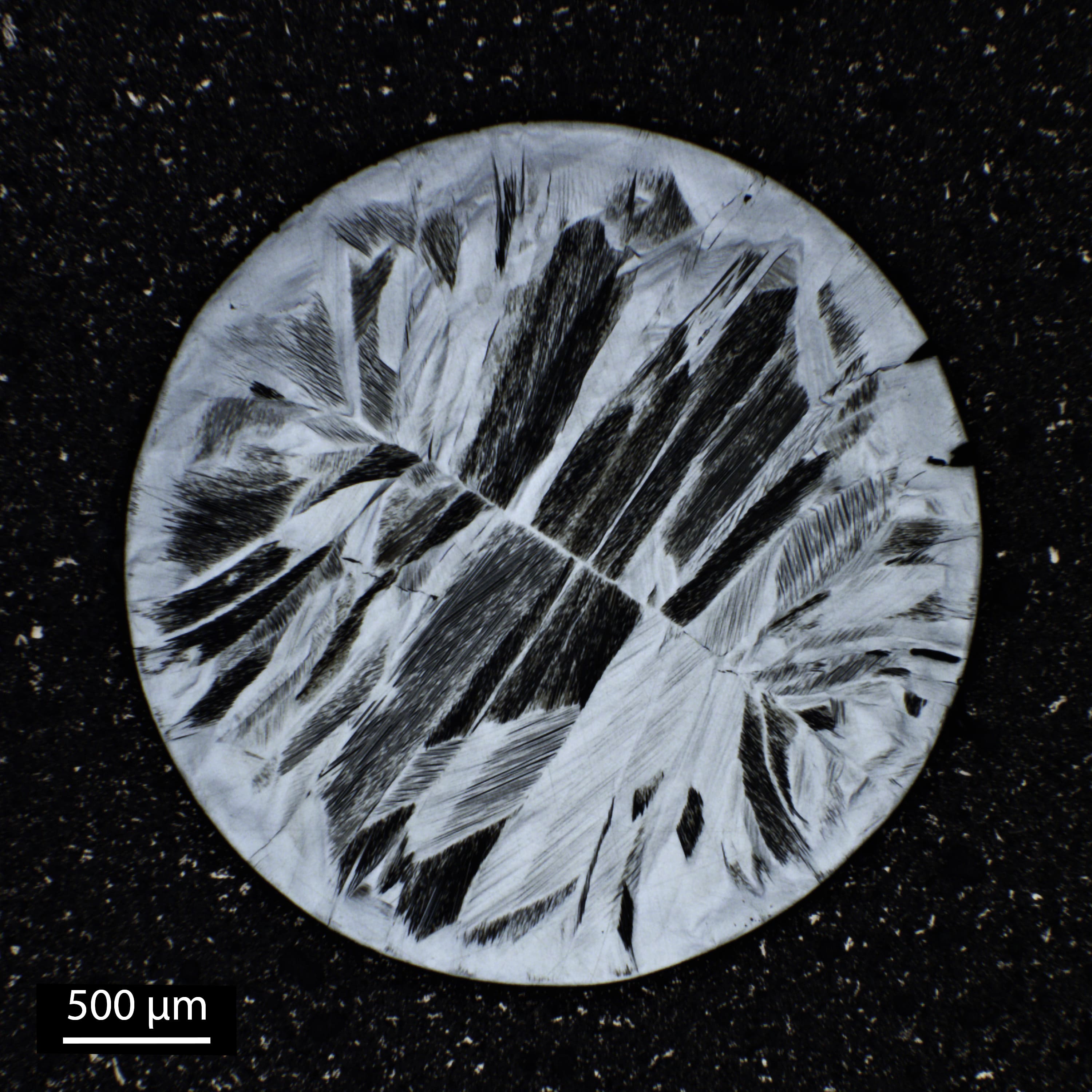
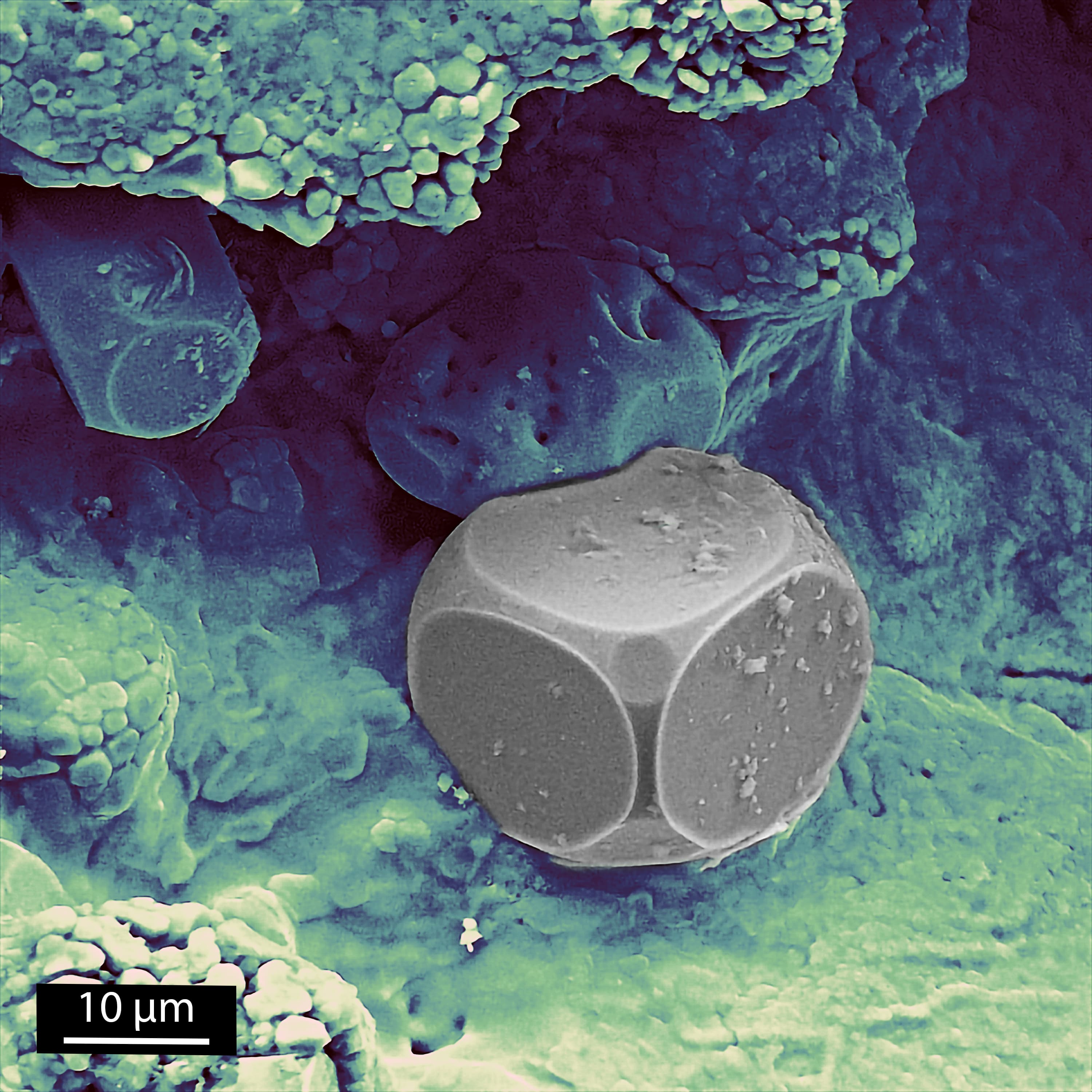
Suction-cast Ni-Mn-In exhibiting thermally-induced martensite adapted to radially symmetric grains. The cylinder has a diameter of 3 mm.
Kerr microscopy from a Nd-Fe-Ti sample after high temperature annealing.
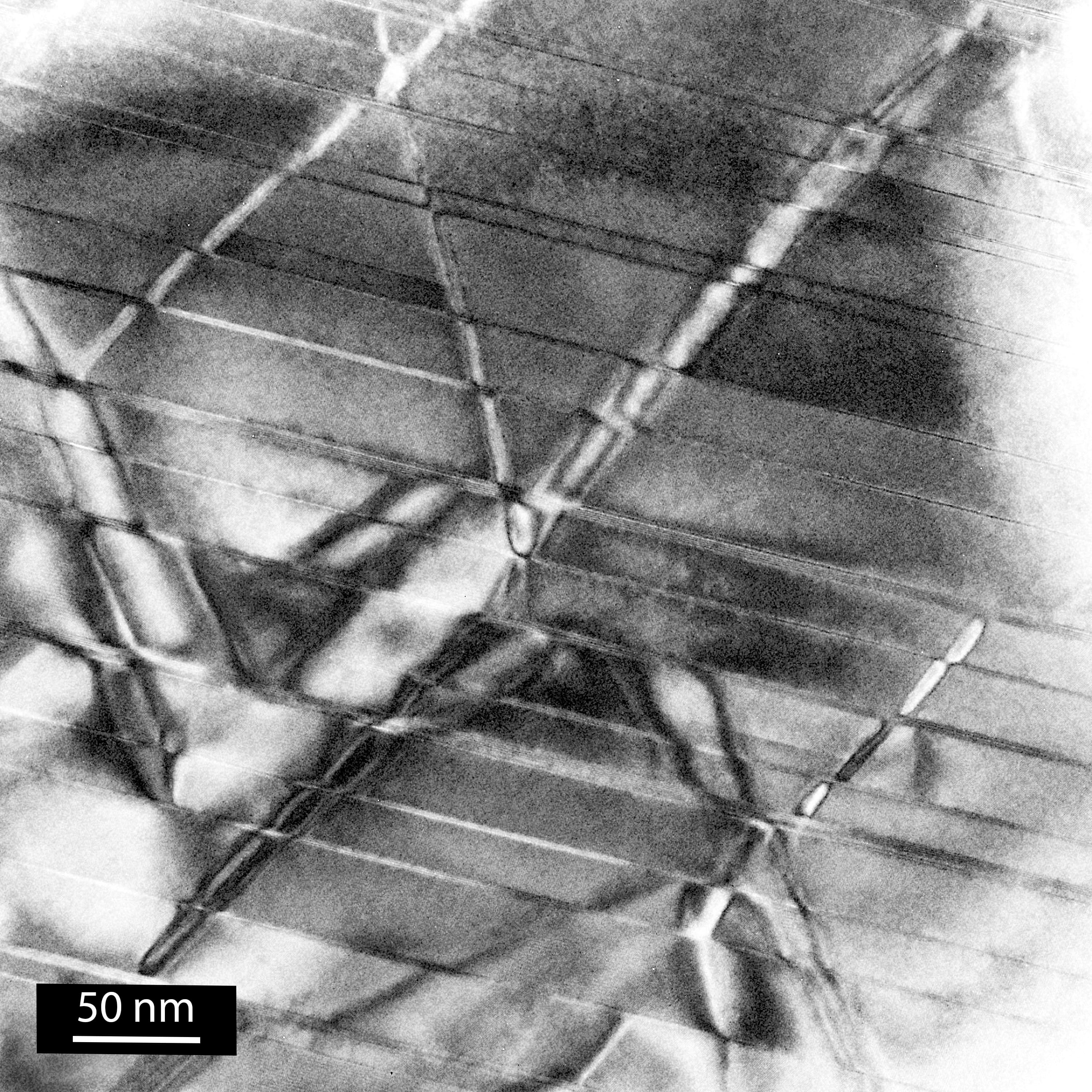
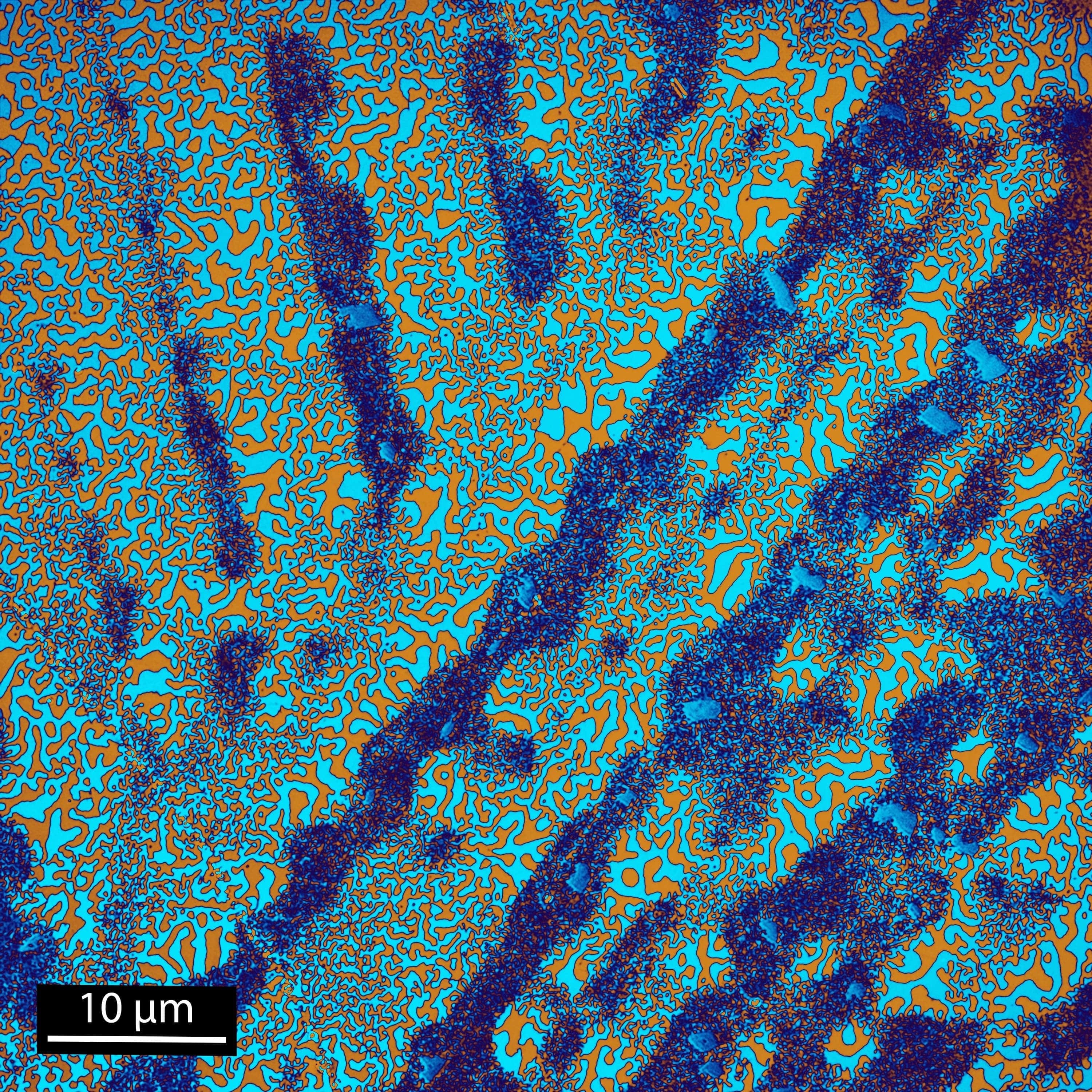
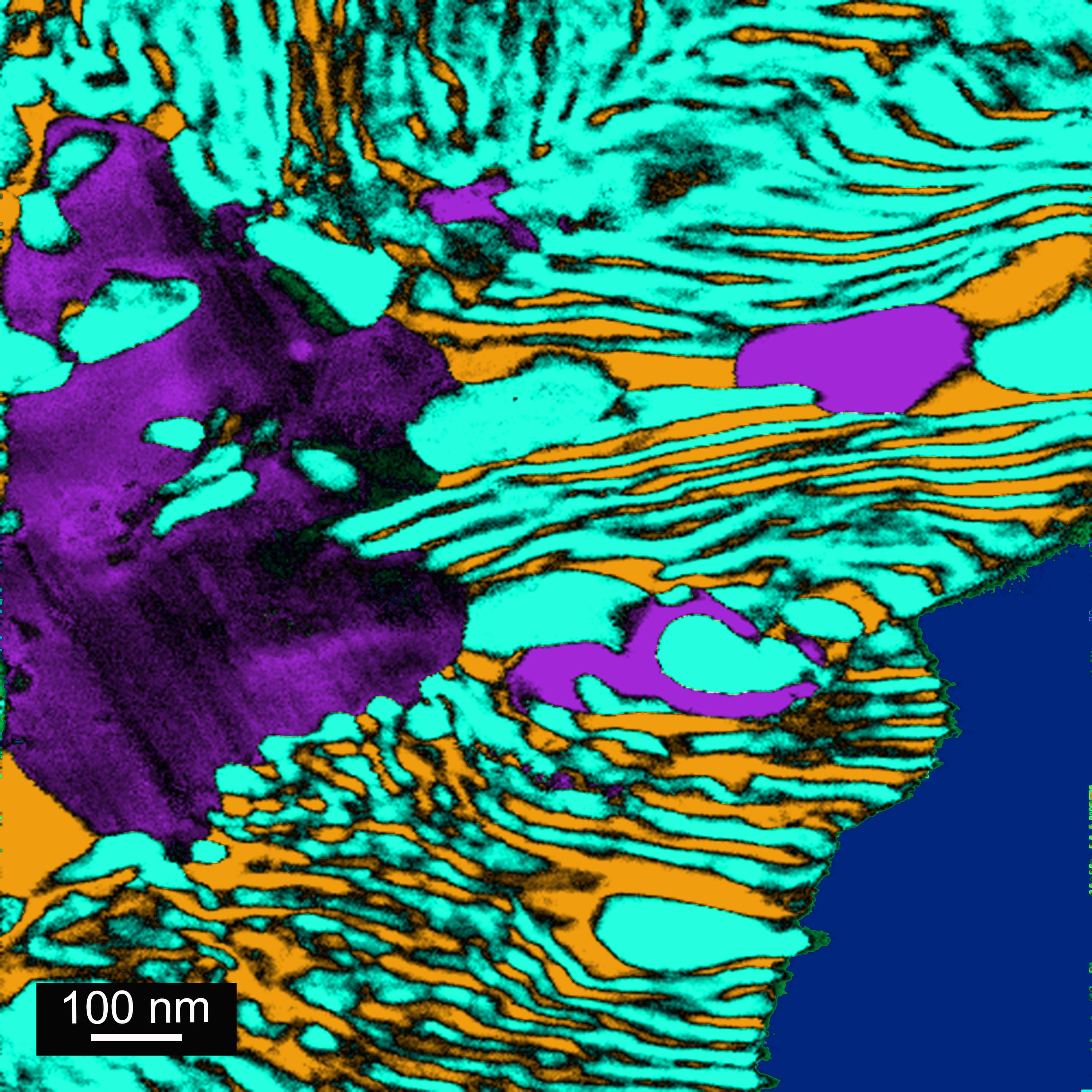
Bright field TEM image of a high temperature Sm-Co permanent magnet, showing the Z-phase platelets, SmCo5 diamond-shaped cellular structure and the Sm2Co17 matrix.
SEM image of a NiMnInCo Heusler alloy showing martensite variants.

Winter Day in Magnetic Forest (Winner of 2022 Joint MMM-Intermag Conference Art in Magnetis Showcase) The motion of Superparamagnetic iron oxide nanoparticles (SPIONs) dispersed in water which are drop casted and sandwiched between two lamellas. With the help of external magnetic field forest like features are observed with an optical microscope. The image has approximate dimensions of 1000*500 µm2.
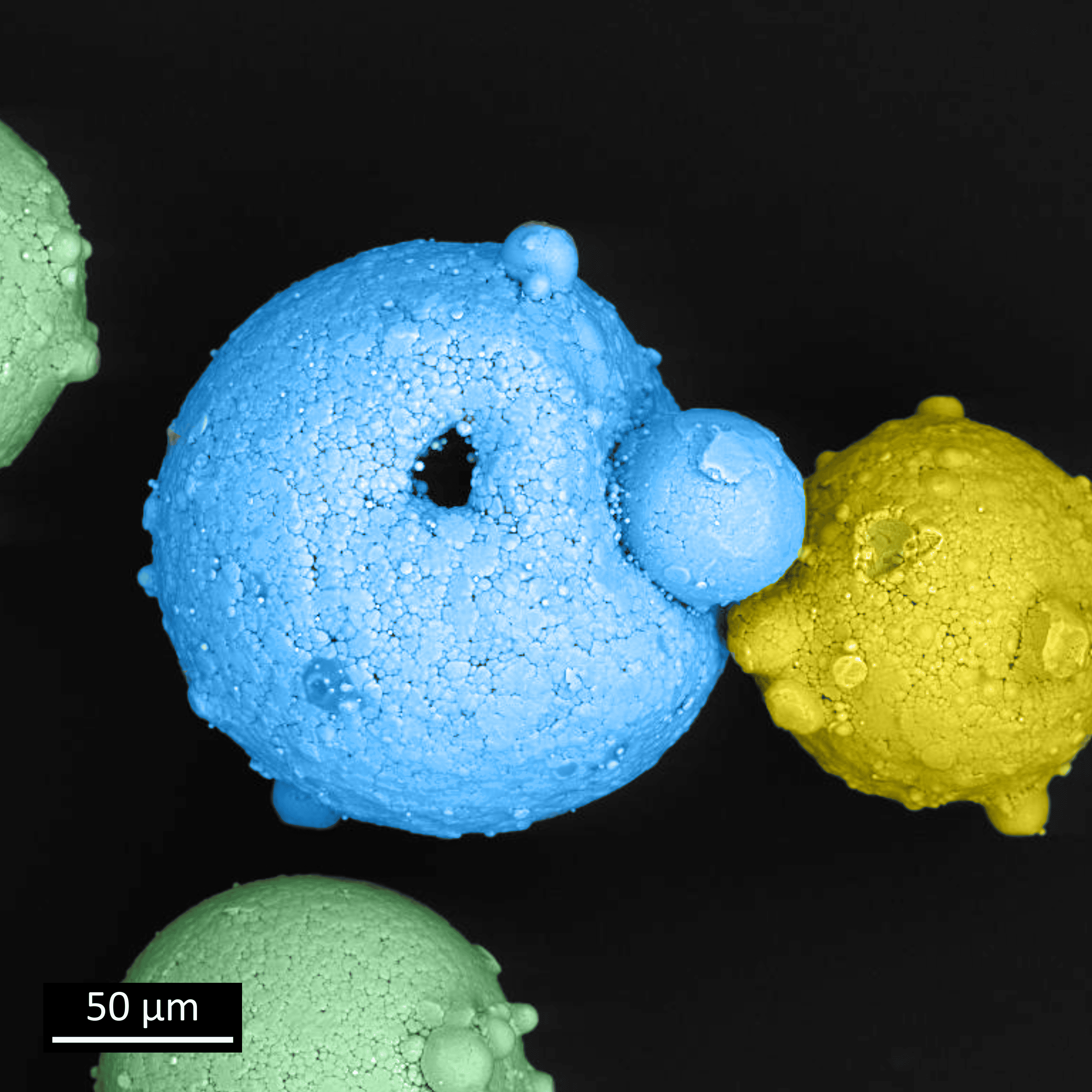
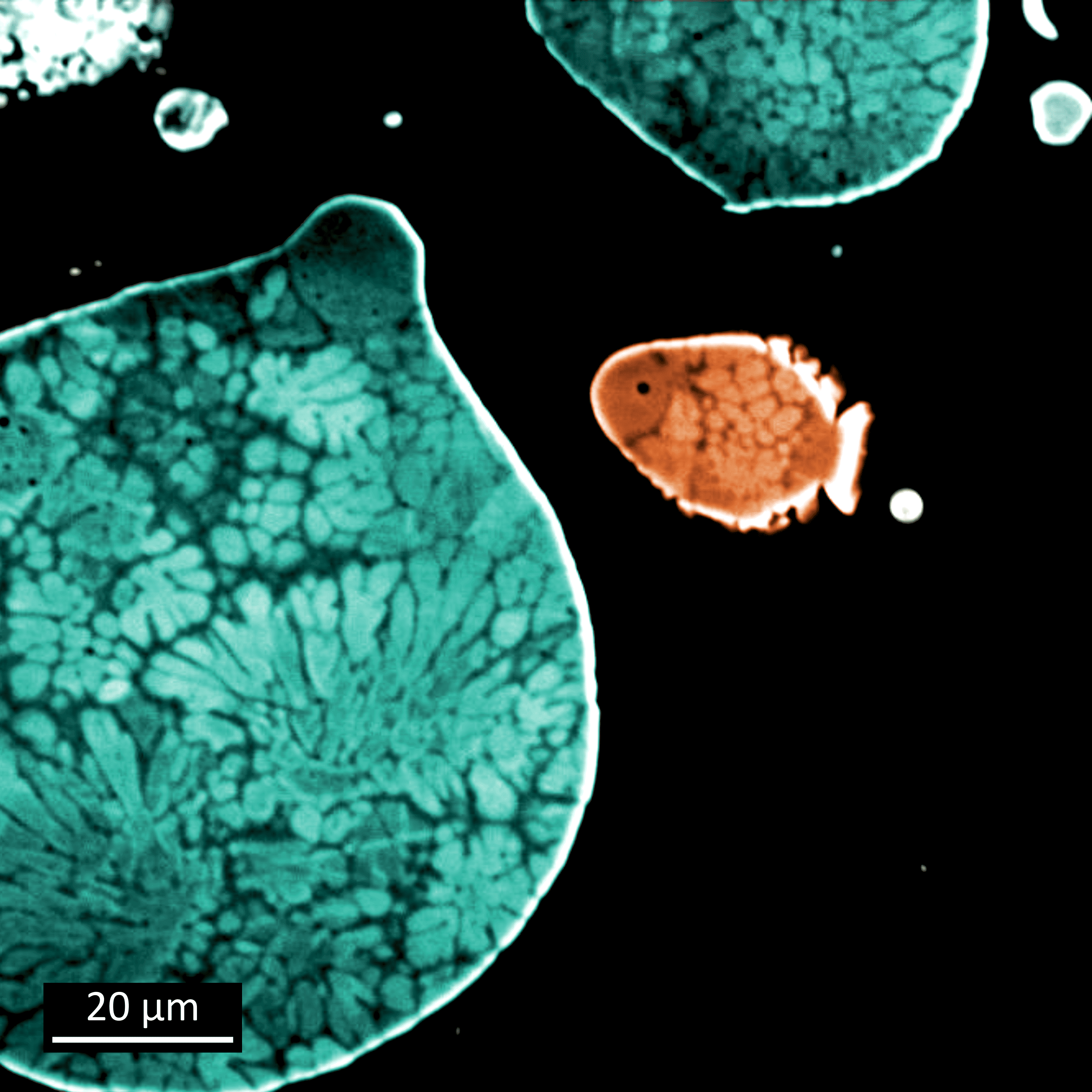
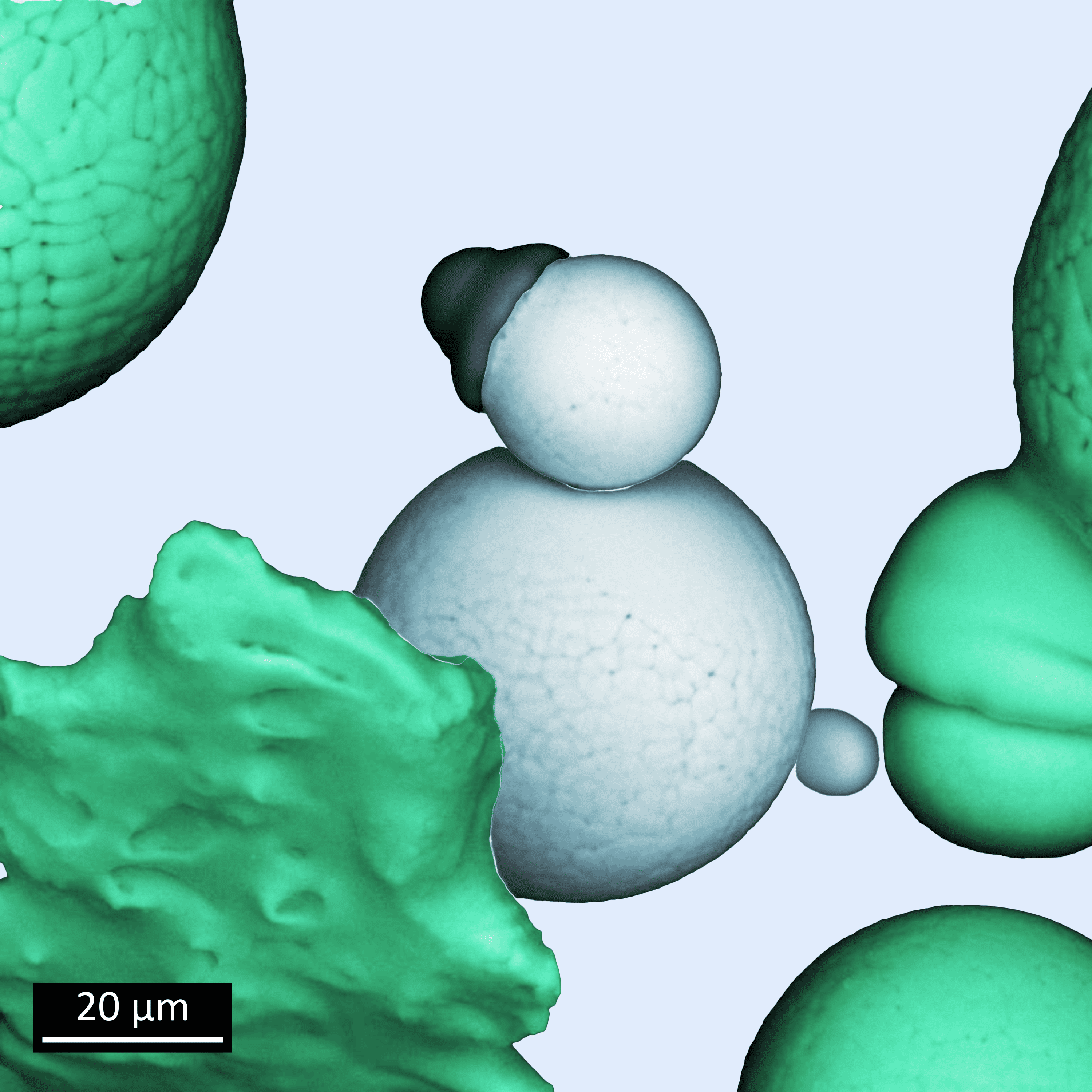
Parent and sleeping child: Two Ni-Mn-Sn particles. The spherical powder was prepared by gas-atomization and act as a base material for additive manufacturing of ferromagnetic Ni-Mn-Sn shape memory alloy.
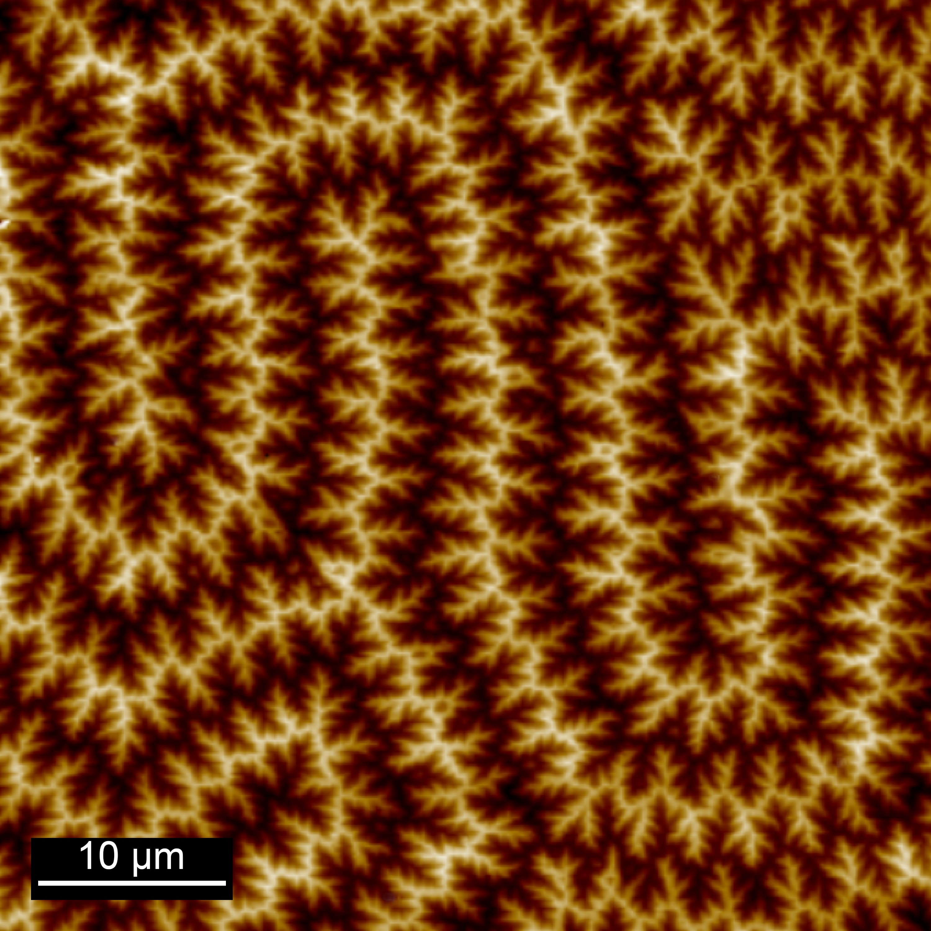
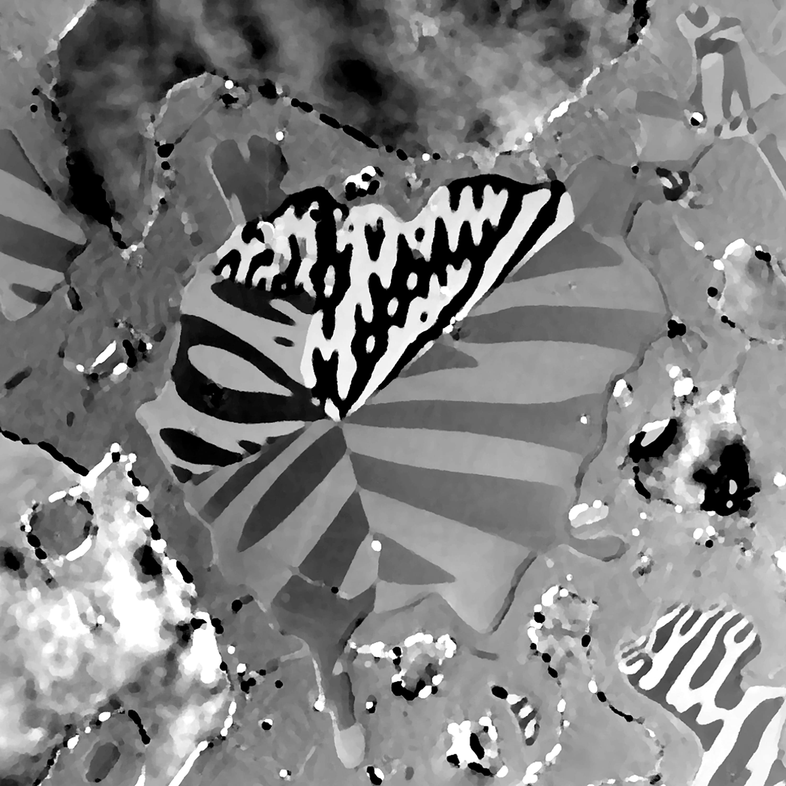
Magnetic force microscopy of Fe3Sn2 revealing uniaxial anisotropy through the magnetic domain structure analysis.
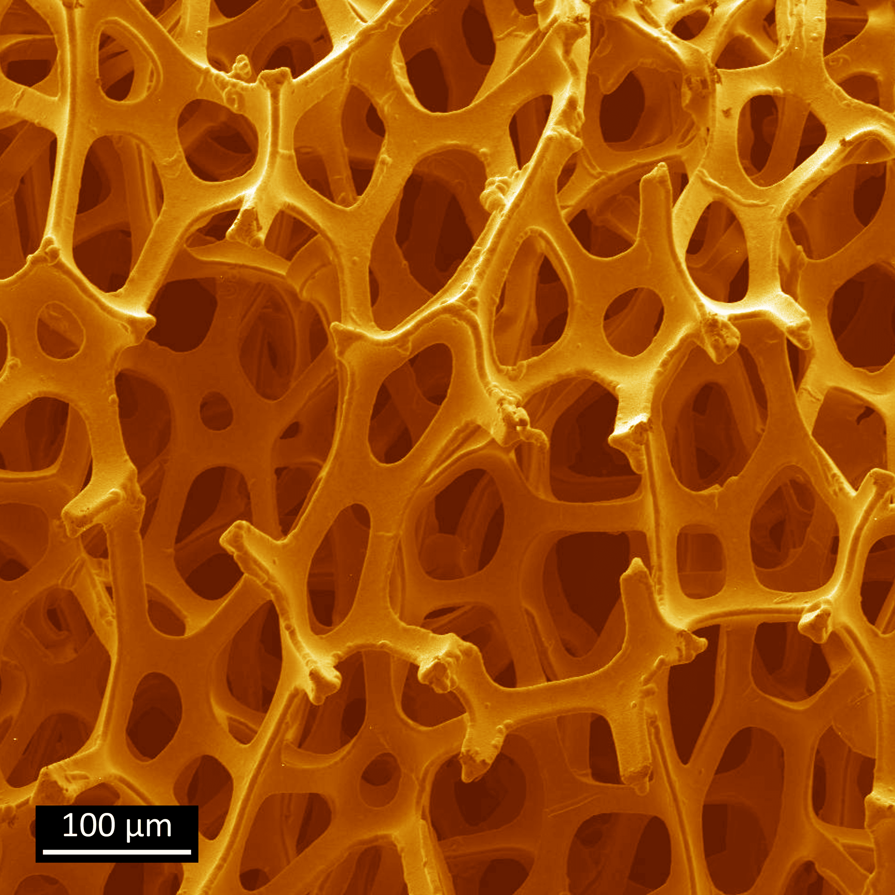
Nickel Foam: The Versatile Beauty - Featuring a Unique Interlinked Three-Dimensional Structure, High Porosity, Excellent Conductivity, and Chemical Resistance. Backbone and Active Material for a Wide Range of Electrochemical Applications, Including Batteries, Sensors, Supercapacitors, and Electrolysis.

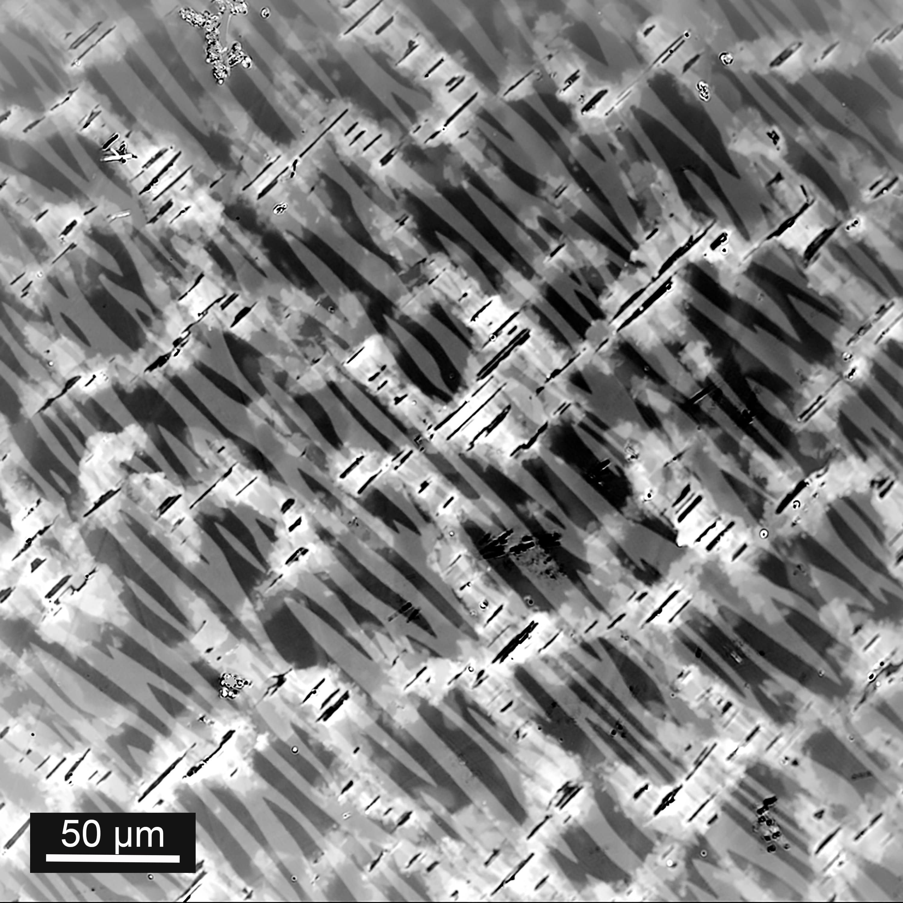
Backscattered electron image of a NiMnInCo Heusler alloy showing Co-rich face-centered cubic γ precipitates after annealing.
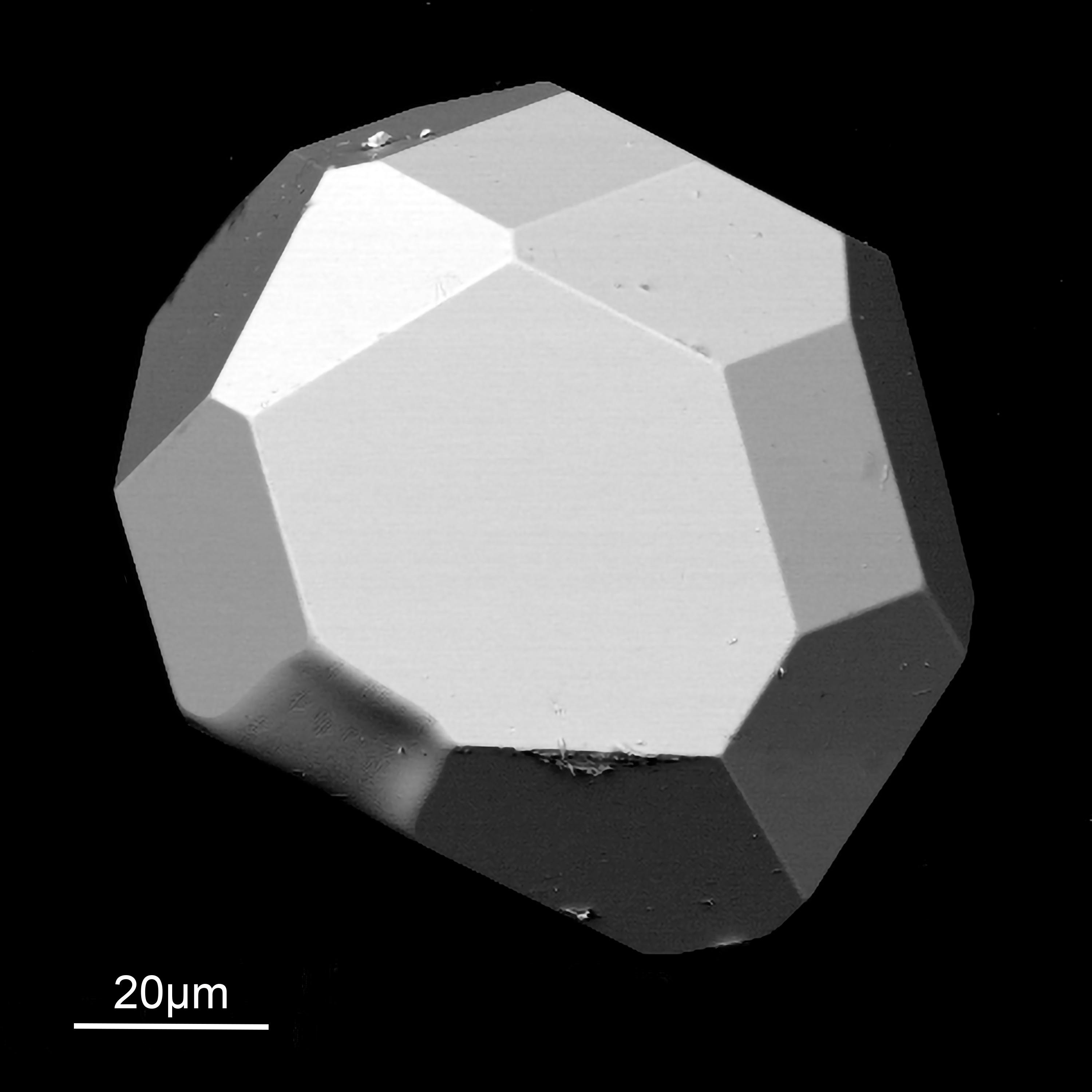
Mn3GaC single crystal grown on a Ga-rich secondary phase formed by solid-state reaction.
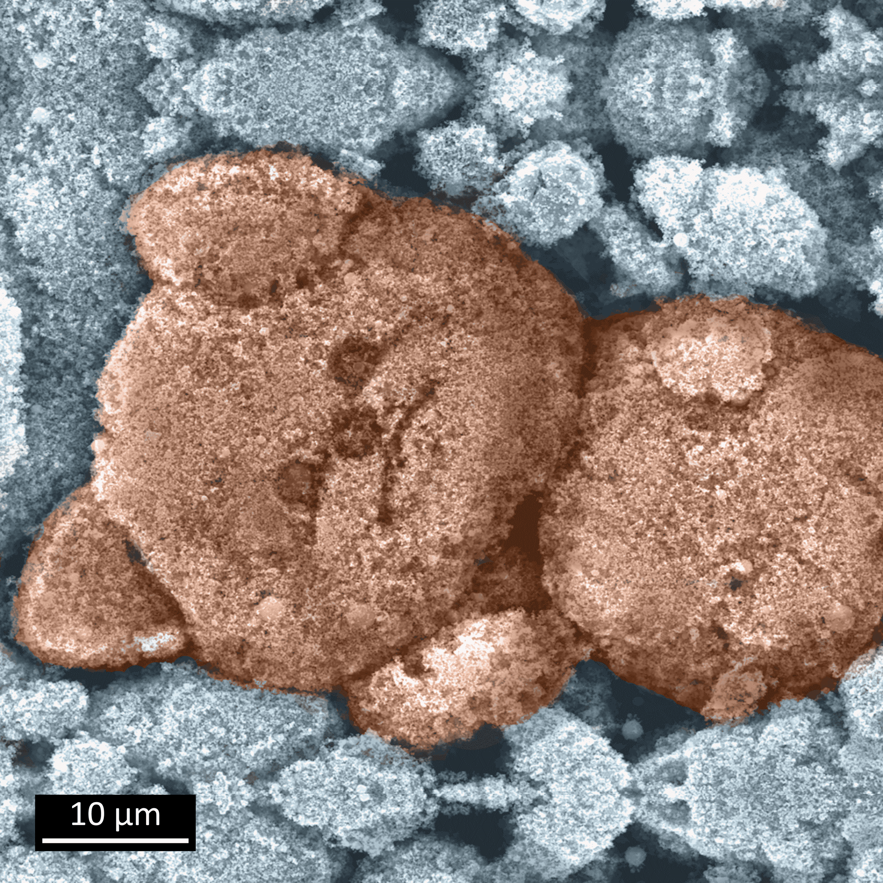
Nanoparticle Feddy Bear (Finalist of INTERMAG 2021 Magnetism as Art Showcase): This naturally-formed teddy bear consists of iron nanoparticles obtained by hydrogen reduction of Fe2O3 . The average particle size is about 50 nm and they form large micrometer-range clusters. The nanoparticles possess high saturation magnetization and a coercivity that exceeds the theoretical anisotropy field value.
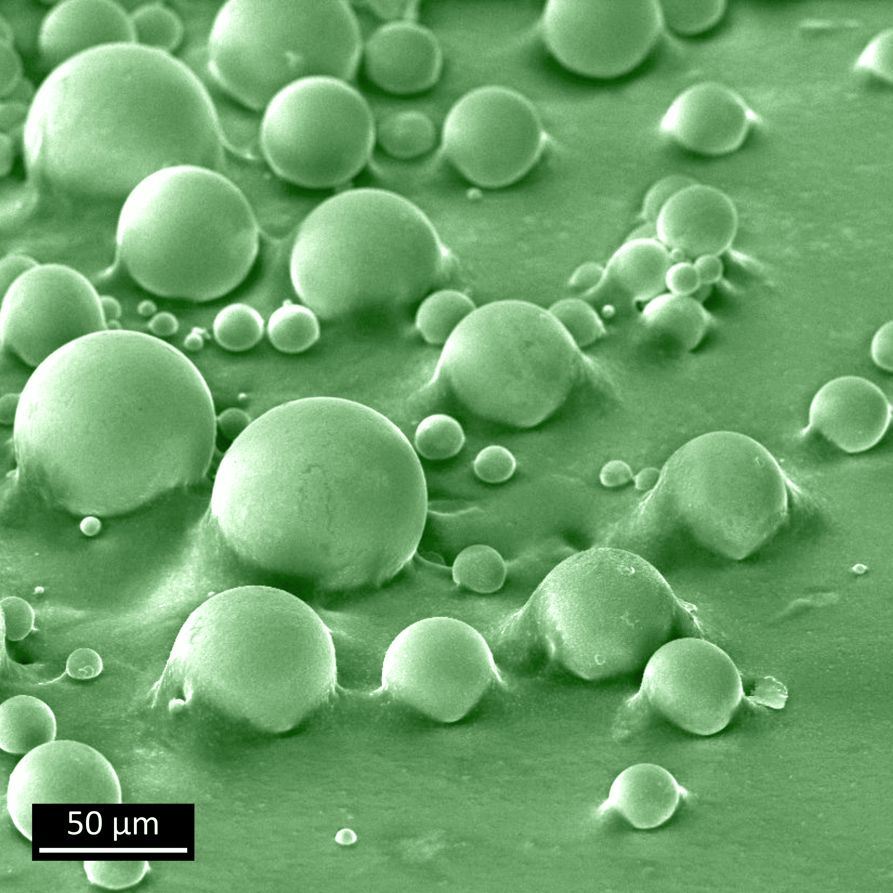
Stuck in a moment: Hard magnetic NdFeB microparticles produced by gas atomization. Such particles are well-suited for additive manufacturing of permanent magnets because their spherical shape ensures good flowability. These NdFeB microparticles are ‘stuck in a moment’ (actually stuck on a carbon tape) and can't get out of it
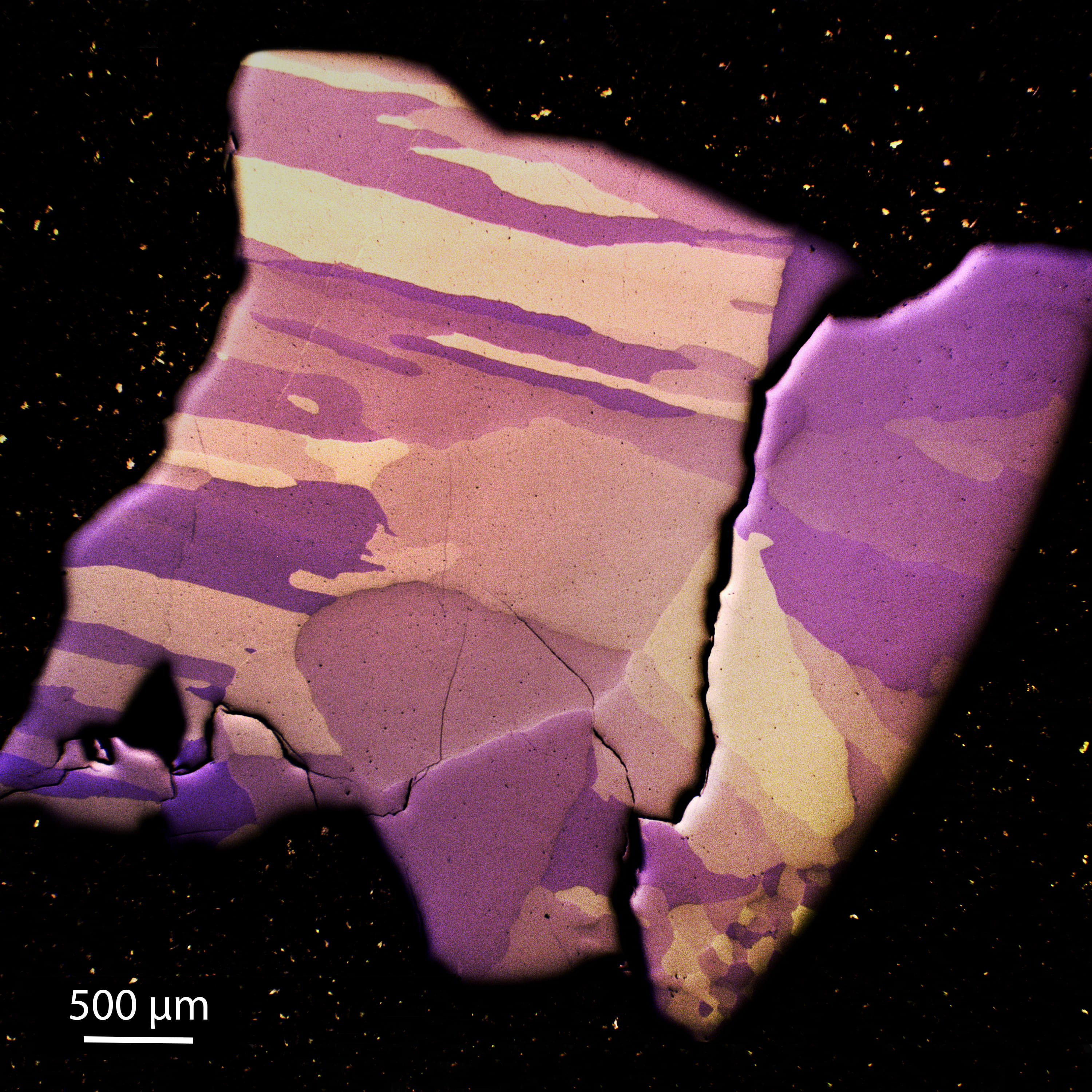
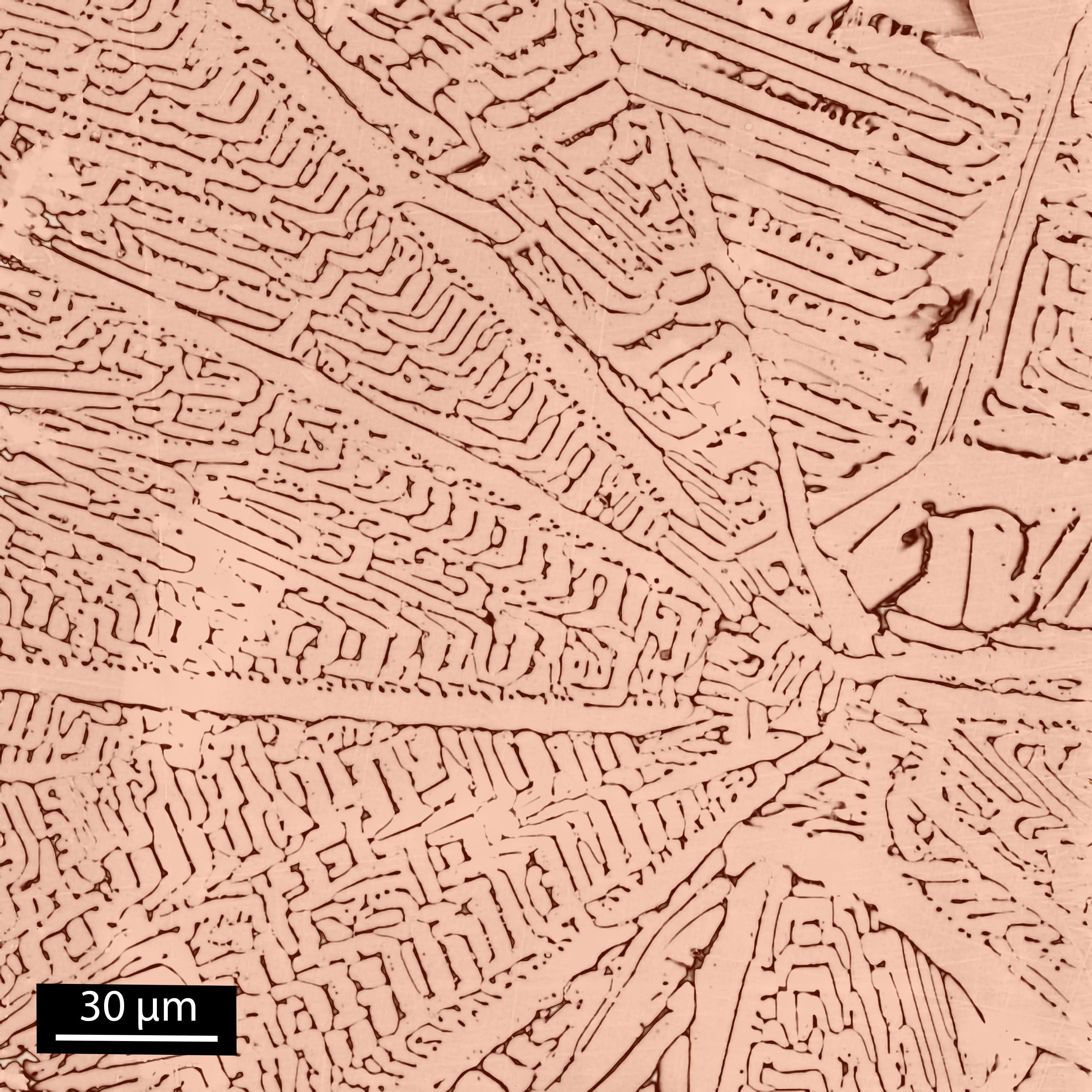
Grain structure of an arc molten FeNiSi particle visualized by optical microscopy using polarized light (particle size about 2.5 mm).
Colored Kerr image from a commercial SmZrCoFeCu alloy after precipitation hardening annealing.
The backscattered electron image (Winner of the Magnetism as Art Showcase of MMM 2022) shows the cross-section of Ni-Mn-Sn powder processed by gas-atomization.
Gas atomized Mn-Al-C materials where different cooling rates result in different morphology.
Videos
Martensitic transition of Heusler alloys
Phase transition of Ni37Co13Mn34Ti16 Heusler alloy from austenite (high temperature) to martensite (low temperature state) by temperature-dependent in-situ optical polarization microscopy.

Magnetic-field induced phase transition of Heusler alloys
Magnetic-field induced phase transition of Ni37Co13Mn34Ti16 Heusler alloy from martensite to austenite by applying a magnetic field from 0 to 1 T at a constant temperature of 250.5 K using magnetic-field-dependent in-situ optical polarization microscopy.

Active magnetocaloric regenerator
Schematic of an active magnetocaloric regenerator for caloric cooling applications showing the contributions of lattice and magnetic spin subsystem during the cycle.

Gauss canon in slow motion
Acceleration of steel balls using strong Nd-Fe-B magnets in a “Gauss canon” arrangement (shown in slow motion).

Hydrogen Decrepitation of as-cast NdFeB
Hydrogen Decrepitation (HD) of as-cast NdFeB at 1 bar and room temperature, a common process in the processing chain for high performance magnet production (accelerated x4).

Hydrogen Decrepitation of a sintered NdFeB magnet
Hydrogen Decrepitation (HD) of a sintered and coated NdFeB magnet at 1 bar and room temperature, an efficient process for functional recycling of scrap magnets from E-motors, hard disk drives, and wind turbines (accelerated x90).

Contact
Prof. Dr. Oliver Gutfleisch

oliver.gutfleisch@tu-...
work +49 6151 16-22150
fax +49 6151 16-22145
Work
L2|07 111
Peter-Grünberg-Str. 16
64287
Darmstadt